Case Report
Volume 4 Issue 1 - 2020
Treatment with Locking Plate Fixation for Distal End Fractures of the Clavicle
Department of Orthopaedic Surgery, Jikei University School of Medicine.
*Corresponding Author: Mamoru Yoshida, MD, PhD. Associate professor, Department of Orthopaedic Surgery, Jikei University School of Medicine, Nishi-shinbashi, Minato-ku, Tokyo, 105-8461 Japan.
Abstract
We conducted surgical treatment with newly designed locking plates for distal end fractures of their clavicles in 30 patients (18 male and 12 female). The average age of patients was 55 years (range 18 to 81 years) and the mean follow-up duration was 17 months (range 13 to 24 months). There were 3 type I, 4 type IIA, 13 type IIB, 4 type V and 6 type VI fractures using the Craig-Takubo classification system. We used locking plates with or without distal wiring followed by active or passive exercises for a range of motion in shoulders for all patients. All fractures united without any adverse effects and all devices were surgically removed 10 to 12 months after surgery. No significant differences were observed for the UCLA shoulder rating score between the dominant side and the opposite healthy side. Surgical fixation with the newly designed locking plates was reliable and safe for the treatment of the distal end fractures of clavicles without surgical reconstructions of coracoclavicular ligaments.
Keywords: Distal end fracture of the clavicle; locking plate fixation
Introduction
For the optimal management of distal end fracture of the clavicle, various treatment modalities are available but no consensus has been reached. A surgical therapy with a hook plate complicated the severe erosion or the fracture of the acromion [1-3] and fixation of the trans-acromioclavicular joint with tension band wiring or other devices restricted the range of scapula-thoracic motion in shoulders [4-7]. We attempted to use newly designed and precontoured locking plates, for which multiple small locking screws stably fixed the distal end fragment of the clavicle, for distal end fractures of the clavicle.
Case Report
We treated 30 patients with distal end fractures using locking plates (HOMS Engineering, Nagano, Japan). There were 18 men and 12 women and the average age of patients was 55 years (range 18-81 years). Average time interval between the initial injury and surgery was 6.3 days (range 3-12 days) and mean follow-up duration was 17 months (range 13-24 months). We examined plain X-ray images or computed tomography to diagnose and classify the fracture type. There were 3 type I, 4 type IIa, 13 type IIb, 4 type V and 6 type VI using the Craig-Takubo classification system. All surgeries were performed by the first author under general anesthesia. The plate was fixed with 3 locking screws or conventional cortical screws 3.5 mm in diameter at the medial end and 3 to 6 mini locking screws 2.7 mm in diameter at the distal end of the clavicle. Additionally the distal end fragments were fixed with or without a wiring system using 1 or 2 cables. Positions for the placement of the locking plates and ideal lengths for the screws were confirmed by fluoroscopy. Abduction pillow braces were used to support the clavicle after surgery and active or passive exercises for a range of motion in shoulders were started 2 or 3 days after surgery. The time to union of the fracture was assessed by examining plain X-ray images or computed tomography. All plates, screws and wiring cables were surgically removed 10 to 12 months after first internal fixation surgery and functional outcomes were assessed 1 to 2 months after second surgery to remove the instrumentation.
All fractures united without adverse effects and the average time to union of the fracture was 15.6 weeks (range 12-18 weeks). There was no re-fracture of any clavicle after the second surgery to remove instrumentation. No significant differences were observed for the range of motion of shoulders, which were elevation, abduction, external rotation and internal rotation between the dominant side and the opposite healthy side at 1 to 2 months after the second surgery. The Japanese Orthopedic Association mean shoulder score was 98 points and no significant differences were observed for the UCLA shoulder rating score between the dominant side and the opposite healthy side at 1 to 2 months after the second surgery. There were a mean 4.2 distal mini locking screws (range 3-6) and a mean 1.0 cable (range 0-2).
Case 1:
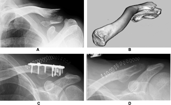
A 38-years-old, man with a Craig-Takubo type V fracture. We used 6 distal locking screws and 2 cables. The distal end fracture united and all devices were removed 10 months after surgery (Fig. 1). No significant difference in UCLA shoulder rating score was observed between the dominant side and the opposite healthy side at 1 month after the second surgery to remove the instrumentation.
A 38-years-old, man with a Craig-Takubo type V fracture. We used 6 distal locking screws and 2 cables. The distal end fracture united and all devices were removed 10 months after surgery (Fig. 1). No significant difference in UCLA shoulder rating score was observed between the dominant side and the opposite healthy side at 1 month after the second surgery to remove the instrumentation.

Figure 1: Case 1. A 38-years-old, man. A. A plain X-ray image showing the anterior-posterior view of the Craig-Takubo type V fracture of the clavicle. B. 3D image of the distal end fracture of the clavicle reconstructed using computed tomography. C. A plain X-ray image of the anterior-posterior view of the clavicle fixed with the locking plate after surgery. Six mini distal locking screws and 2 cables fixed the distal end fragment of the clavicle. D. A plain X-ray image of the anterior-posterior view of the clavicle after removal of all devices. The distal end of the fracture united and no deformity was observed.
Case 2:
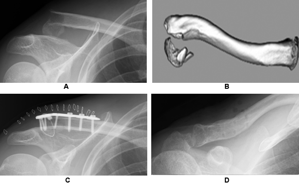
A 43-years-old, man with a Craig-Takubo type VI fracture. We used 3 distal locking screws and 1 cable. The distal end fracture united and all devices were removed 12 months after surgery (Fig. 2). No significant difference in UCLA shoulder rating score was observed between the dominant side and the opposite healthy side 1 month after the second surgery to remove the instrumentation.
A 43-years-old, man with a Craig-Takubo type VI fracture. We used 3 distal locking screws and 1 cable. The distal end fracture united and all devices were removed 12 months after surgery (Fig. 2). No significant difference in UCLA shoulder rating score was observed between the dominant side and the opposite healthy side 1 month after the second surgery to remove the instrumentation.

Figure 2: Case 2. A 43-years-old, man. A. A plain X-ray image showing the anterior-posterior view of the Craig-Takubo type VI fracture of the clavicle. B. 3D image of the distal end fracture of the clavicle reconstructed using computed tomography. C. A plain X-ray image of the anterior-posterior view of the clavicle fixed with a locking plate after surgery. Three mini distal locking screws and 1 cable fixed the distal end fragment of the clavicle. D. A plain X-ray image of the anterior-posterior view of the clavicle after removal of all devices. The distal end of the fracture united and no deformity was observed.
Discussion
The present study demonstrated that the displaced distal clavicle fractures treated with the distal locking plate with or without cable fixation accomplished good results in terms of both a bony union and a functional outcome with rarely any complications compared with those of other treatment modalities. These results indicated that the fixation of the distal end fragment of clavicle fractures with multiple mini locking screws was stable and suitable for allowing early mobilization or exercise. Various types of locking plates have been released by various manufactures [8-12]. Compared with other types of locking plates, the locking plates used in the present study had several advantages in that the shape of the plate is precontoured and anatomically matched with the shape of the clavicle, the plate is thinner than other plates and the distance between the distal end of the plate and the distal screw hole is the shortest of all commercially available locking plates. These advantages contributed to the precise placement of the plate and minimized stimulation or mechanical stress against the soft tissues around the clavicle and interference of acromioclavicular joints. We consider that these factors allowed stable fixation of the distal end fractures of the clavicles, even though the bony size or quality of the distal end fragment was small or poor, and did not restrict the range of motion in shoulders or cause any adverse effects such as skin incision problems after surgery.
The necessity of the cable system is controversial. The fracture united without the cable system with fixation by 3 to 6 mini locking screws in 5 cases, which were 2 type I and 3 type II fractures, in the present study. However, there were a few failure cases without the cable system with fixation by 3 mini locking screws in elder osteoporotic patients in other institutes. It is estimated that the cable system is necessary to prevent the initial failure in the case of elder osteoporotic patients and of fixation by less than 3 screws. Further investigations are necessary to confirm the reliability or availability of this surgical modality and to determine the necessity of the cable system fixed to the distal end because the number of cases in the present study is small and may not provide sufficient power to determine differences.
Conclusion
Surgical fixation with the newly designed locking plates was reliable and safe for the treatment of the distal end fractures of clavicles without surgical reconstructions of coracoclavicular ligaments.
Acknowledgements
Funding acknowledgements
Funding acknowledgements
Declaration of Conflict of Interest
None
None
References
- Flinkkila T., et al. “Hook-plate fixation of unstable lateral clavicle fractures: a report on 63 patients”. Acta Orthopaedica 77.4(2006): 644-649.
- Kashii M., et al. “Surgical treatment of distal clavicle fractures using the clavicular hook plate”. Clinical Orthopaedics and Related Research 447(2006):158-164.
- Haidar SG., et al. “Hook plate fixation for type II fractures of the lateral end of the clavicle”. Journal of Shoulder and Elbow Surgery 15.4(2006): 419-423.
- Fann CY., et al. “Trans-acromial Knowles pin in the treatment of Neer type 2 distal clavicle fractures: A prospective evaluation of 32 cases”. The Journal of Trauma and Acute Care Surgery 56.5(2004):1102-1105.
- Badhe SP., et al. “Tension band suturing for the treatment of displaced type-2 lateral end clavicle fractures”. Archives of Orthopaedic and Trauma Surgery 127.1 (2007): 25-28.
- Jou IM., et al. “Treatment of unstable distal clavicle fracture with Knowles pin”. Journal of Shoulder and Elbow Surgery 20.3(2011): 414-419.
- Samy M and Khanfour A. “Extra-articular fixation of displaced fracture lateral end clavicle”. European Journal of Orthopaedic Surgery & Traumatology 21.8(2011): 557-561.
- Anderson JR., et al. “Precontoured superior locked plating of distal clavicle fractures: a new strategy”. Clinical Orthopaedics and Related Research 469.12(2011): 3344-3350.
- Lee SK., et al. “Precontoured locking plate fixation for displaced lateral clavicle fractures”. Orthopedics 36.6(2013): 801-807.
- Zhang C., et al. “Comparison of the efficiency of a distal clavicular locking plate versus clavicular hook plate in the treatment of unstable distal clavicle fractures and a systematic literature review”. International Orthopaedics 38.7(2014): 1461-1468.
- Choo SK., et al. “The surgical outcome of unstable distal clavicle fractures treated with 2.4 mm volar distal radius locking plate”. Journal of the Korean Fracture Society 28.1(2015): 38-45.
- Vaishya R., et al. “Outcome of distal end clavicle fractures treated with locking plates”. Chinese Journal of Traumatology 20.1(2017): 45-48.
Citation: Mamoru Yoshida., et al. “Treatment with Locking Plate Fixation for Distal End Fractures of the Clavicle”. Orthopaedic Surgery and Traumatology 4.1 (2020): 13-16.
Copyright: © 2020 Mamoru Yoshida., et al. This is an open-access article distributed under the terms of the Creative Commons Attribution License, which permits unrestricted use, distribution, and reproduction in any medium, provided the original author and source are credited.


 Scientia Ricerca is licensed and content of this site is available under a Creative Commons Attribution 4.0 International License.