Case Report
Volume 1 Issue 3 - 2017
The Tillaux Fracture in Adult
Department of Orthopaedics, NMC Specialty Hospital Abudhabi
*Corresponding Author: Dr. Surendra Shetty, Department of Orthopaedics, NMC Specialty Hospital Abudhabi.
Received: January 20, 2017; Published: January 25, 2017
Introduction
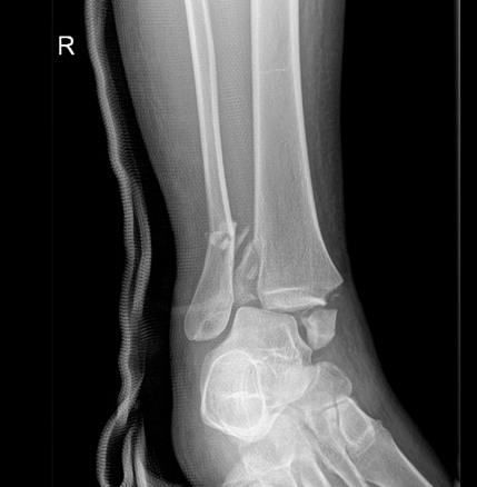
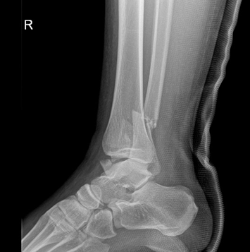
We describe a bimalleolar fracture of the right ankle in a 24 year old man with an associated ankle subluxation and fracture of the anterolateral aspect of distal tibia - the adult tillaux fracture. Adult tillaux fracture is an extremely rare fracture pattern with very few cases reported in the literature.
Summary
The case described is of a 24 year old male who sustained the injury while trekking in the hills. Careful evaluation of x rays AP, lateral and oblique views are helpful in identifying the tillaux fracture. We would also recommend evaluation with CT.
Case Presentation
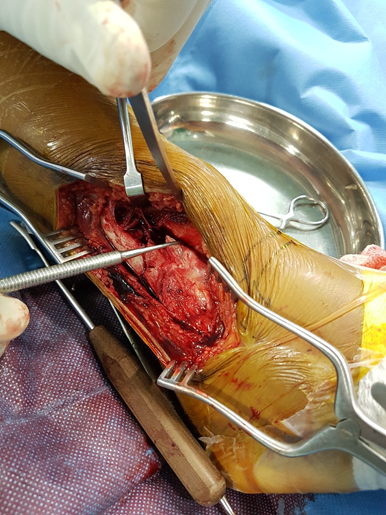
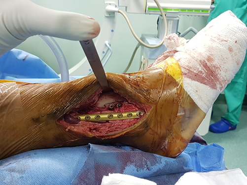
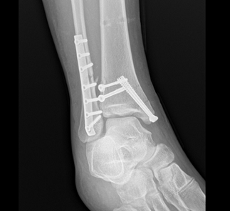
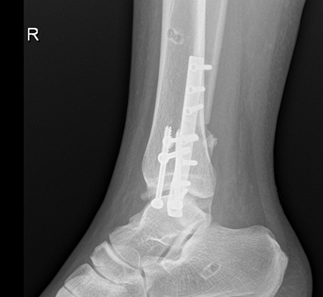
The case was seen in emergency 12 hours after the fall. He presented with a swollen ankle and foot. Skin overlying the medial malleolus had abrasions and gross ecchymosis. Distal neurovascular status was intact. X rays showed fracture of the lower fibula, medial malleolar fracture with separation of the malleolus, subluxation of talus with disruption of the ankle joint and fracture of the antero lateral aspect of distal tibia. The patient was taken to operating room. Under GA the ankle was reduced and external fixator was applied with pins in calcaneum and tibia. CT scan confirmed the finer details of the anterolateral fracture pattern of distal fibula. After 10 days when skin condition improved and swelling reduced, definitive open reduction and internal fixation was done for the fractures. The medial malleolus was fixed with open reduction and internal fixation with two 4.5 mm cannulated screws. Next the fibula fracture was exposed with a lateral incision. Through the same incision, the anterolateral aspect of tibia was exposed extra periosteally and the tillaux fragment was fixed with one cannulated 4.5 mm screw and one 3.5 mm screw proximal towards apex of the fracture. Fibula frature was fixed with one third tubular plate and 3.5 mm screws. The syndesmosis was assessed and found to be stable.
Discussion
At the lower end of tibia, about 5–6 cm above the ankle joint line, there is a concave shallow triangle facing laterally to receive distal shaft of fibula. At the inferior border of this depression there are anterior and posterior tibial tubercles. The interosseous ligament attaches to tibia in this groove and to a similar area on the adjacent fibular shaft forming a syndesmosis. The antero-inferior tibiofibular ligament attaches to the anterior tubercle and the anterior aspect of the lateral malleolus and the postero- inferior tibiofibular ligament attaches to the posterior tibial tubercle on the posterior aspect of the lateral malleolus.1
The distal tibia and fibula is stabilized with four ligaments. The anterior and posterior tibofibular ligaments, transverse tibiofibular ligament and interosseous membrane. Tillaux fracture represents avulsion of antero-lateral tibial epiphysis due to the strong pull of tibio-fibular ligament. This happens usually in external rotation injuries. This is common in adolescents and rare in adults. In adults, because the physis is completely closed and the bone is stronger than the ligaments, the latter gives way instead of avulsing the tibial fragment from its attachment, resulting in a ligament injury known as a Tillaux lesion. However in rare cases, the distal tibial tubercle is avulsed off the anterolateral aspect of the distal tibia resulting in Tillaux fracture.
In adult type of tillaux fracture, the avulsed fracture fragment is triangular as compared to the juvenile one, where the fragment is quadrangular.2 Adult pattern of Tillaux fractures are classified into Type A and Type B. Type A is avulsion fractures of the antero-lateral aspect of the distal tibial plafond and Type B is a fracture pattern extending into the medial aspect resulting in antero medial pattern3 If the fracture displacement is less than 2 mm, it can be managed conservatively. If the displacement is more than 2 mm then operative management is required.4 In our case there was difficulty in closed reduction initially in the ER, as the displaced fracture fragment were making the reduction difficult. Even after we achieved reduction the ankle was unstable. Hence we decided to put an external fixator.
References
- Protas JM and Kornblatt BA. “Fractures of the lateral margin of the distal tibia. The Tillaux fracture”. Radiology 138.1 (1981): 55-57.
- Kleiger B and Mankin HJ. “Fracture of the lateral portion of the distal tibial epiphysis”. The Journal of Bone & Joint Surgery 46 (1964): 25-32.
- S Chokkalingam and S Roy. “Adult Tillaux Fractures Of Ankle: Case Report. Adult Tillaux Fractures Of Ankle: Case Report”. The Internet Journal of Orthopedic Surgery 6.1 (2006):
- Sharma B., et al. “The adult Tillaux fracture: one not to miss”. BMJ Case Reports (2013): doi:10.1136/bcr-2013-200105.
Citation:
Surendra Shetty., et al. “The Tillaux Fracture in Adult”. Orthopaedic Surgery and Traumatology 1.3 (2017): 114-117.
Copyright: © 2017 Surendra Shetty., et al. This is an open-access article distributed under the terms of the Creative Commons Attribution License, which permits unrestricted use, distribution, and reproduction in any medium, provided the original author and source are credited.


 Scientia Ricerca is licensed and content of this site is available under a Creative Commons Attribution 4.0 International License.