Research Article
Volume 2 Issue 2 - 2018
Coccidiosis in Domestic Juvenile Pigeons
1Assistant Professor, Department of Veterinary Parasitology
2Assistant Professor, Faculty of Veterinary Medicine, Department of Veterinary Clinical Complex
2Assistant Professor, Faculty of Veterinary Medicine, Department of Veterinary Clinical Complex
*Corresponding Author: S Sivajothi, College of Veterinary Science, Department of Veterinary Parasitology, Proddatur - 516 360, Y.S.R.
Kadapa District, Sri Venkateswara Veterinary University, Andhra Pradesh, India.
Received: April 17, 2018; Published: May 11, 2018
Abstract
During coccidiosis, birds have impaired digestion which leads to passing of watery droppings subsequently loss of body condition in
the individual birds. Sixteen juvenile pigeons were diagnosed that they were suffering with the clinical coccidiosis by the examination
of faecal samples. Flotation technique was adopted for the confirmation of the parasitic oocysts in the faeces of pigeons. All the
pigeons were successfully treated with amprolium.
Keywords: Coccidiosis; Eimeria; Columba livia; Amprolium; Juvenile
Introduction
Different types of internal and external parasites in pigeon’s leads to loss of body condition, severe discomfort and the parasites act as
vectors for other diseases. During the acute stage of disease condition, mortality of the birds can be noticed (Sivajothi). Coccidiosis is one
of the most common and highly pathogenic obligatory enteric protozoans affecting poultry in the world. Juvenile pigeon mortality due to
coccidiosis was varies from 5% to 70% and pigeons between the third to fourth months were highly susceptible (Al-Rubaie). The present
study placed a record on successful treatment of coccidiosis in the juvenile pigeons with amprolium.
Materials and Methods
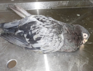
Sixteen birds in a flock of 64 pigeons were identified that they were suffering with watery greenish colour droppings in Proddatur,
YSR Kadapa District of Andhra Pradesh (Figure-1). Faecal droppings were collected in to a sterile container for parasitic ova examination.
Direct microscopic examination was carried out by addition of few drops of distilled water and further samples were examined for the
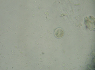
parasitic oocysts by the flotation technique. Microscopic examination of the faecal samples revealed presence of the unsporulated coccidial
oocysts (Figure 2).
Treatment and Discussion
All the birds were treated with oral amprolium (20%) (1 gram of powder in 1 litre of water) solution by addition of the water daily
for seven days. Improvement in the condition of the birds was assessed by the consistency of droppings and activity of the birds. All the
pigeons were recovered uneventfully by the change in the colour and consistency of the faeces without any adverse reactions. After
completion of therapy, multi vitamin syrup was added in the drinking water for nutritional support.
In general, oocysts of Eimeria found in the pigeon feces. It was immature oocysts with colourless, mature oocysts with complete
triple-layered wall and last one is degraded oocyst, which is clark yellow and finely granulated without any refractive granules. Oocysts
are spread by aerosol transmission by means of dust from dried feces or debris from nests or footwear. Juvenile pigeons are anorexic,
dehydrated, dull, emaciated and watery diarrhoea but in four birds, it was hemorrhagic diarrhea. In the present study, diagnosis of the
coccidiosis was done by the examination of the oocysts in the faeces of pigeons. Examination of the coccidian oocysts was enhanced by
the floatation technique. But during the severe pathologic coccidiosis, clinical signs arise before gamogony; therefore, fecal samples will
be negative. Coccidiosis in adult birds is self limiting and treatment is not required but, juvenile pigeons are required treatment. Oocysts
from the effected droppings will become infectious after completion of the sporulation procedure and it will take 1 to 2 days after the
shedding of oocysts from the infected pigeon. Complete eradication of the oocysts in the environment can be achieved by regular removal
of the litter and cleaning the premises with disinfectants (Selvarani).
Amprolium is a pyrimidine derivative, has coccidiostatic properties. It is structurally similar to thiamine and, when ingested by coccidia,
competitively inhibits folic acid metabolism. It is structurally similar to the thiamine and when provided higher dose of amprolium
causes reduction in the thiamine levels further leads to vitamin deficiency to the birds. It is not advisable to give excessive quantity of
thiamine (more than 1:1 ratio of thiamine to amprolium) along with the amprolium therapy which further leads to reduction in the efficacy
of the amprolium towards the coccidiosis in the birds. Toxicity of the amprolium was very minimal due to its poor enteral absorption
and quick elimination from the circulation (Hoskin).
In the previous studies coccidiosis was recorded in different species of the animals and successfully treated with sulphonamide
group of drugs (Reddy). In the present study amprolium was selected to treat the juvenile birds.
Conclusion
Study puts a record on the outbreak of coccidiosis in juvenile pigeons and its successful management with amprolium.
Acknowledgement
The authors are thankful to the authorities of Sri Venkateswara Veterinary University, Tirupati for providing the facilities to carry out the work.
The authors are thankful to the authorities of Sri Venkateswara Veterinary University, Tirupati for providing the facilities to carry out the work.
Conflict of Interest
None.
None.
References
- Al-Rubaie HMA and Mahdii EF “Study the prevalence of pigeon coccidiosis in Baghdad city.” An insight into veterinary education in Iraq 37.1 (2013): 106-108.
- Hoskins JD and Cheney JM. “Parasitology and public health.” Clinical Textbook for veterinary Technicians (1994): 72-95.
- Reddy BS., et al. “Clinical coccidiosis in adult cattle.” Journal of Parasitic Diseases 39.3 (2015): 557-559.
- Selvarani R., et al. “In-vivo evaluation ofanti-coccidial efficacy of Salinomycin and amprolium in commercial chicken.” Indian Journal of Animal Sciences 43.2 (2014): 169 - 173.
- Sivajothi S and Reddy BS. “A Study on the Gastro Intestinal Parasites of Domestic Pigeons in YSR Kadapa District in Andhra Pradesh India.” Journal of Dairy Veterinary & Animal Research 2.6 (2015): 00057.
Citation:
S Sivajothi and B Sudhakara Reddy. “Coccidiosis in Domestic Juvenile Pigeons". Clinical Biotechnology and Microbiology 2.2
(2018): 345-347.
Copyright: © 2018 S Sivajothi and B Sudhakara Reddy. This is an open-access article distributed under the terms of the Creative Commons Attribution License, which permits unrestricted use, distribution, and reproduction in any medium, provided the original author and source are credited.


 Scientia Ricerca is licensed and content of this site is available under a Creative Commons Attribution 4.0 International License.