Case Report
Volume 3 Issue 1 - 2019
Bilateral Panuveitis Revealing Mediterranean Spotted Fever
1Department of Internal medicine. Military Hospital of Gabes. Gabes 6000. Tunisia
2Sfax Faculty of Medicine. University of Sfax. Sfax 3029. Tunisia
2Sfax Faculty of Medicine. University of Sfax. Sfax 3029. Tunisia
*Corresponding Author: Salem Bouomrani, MD, PhD, Department of Internal medicine, Military Hospital of Gabes. Gabes 6000. Tunisia.
Received: January 19, 2019; Published: June 05, 2019
Abstract
Introduction: Mediterranean spotted fever (MSF) is a rickettsial disease caused by the species Rickettsia Conorii, and it can present itself with unexpected and often unusual revealing manifestations that explain the diagnostic difficulties of these infections. We are reporting an original observation of bilateral panuveitis revealing rickettsiosis type MSF.
Case report: A 52-year-old Tunisian patient, with no notable pathological history, was transferred to our department by his city ophthalmologist for the management of acute bilateral non granulomatous panuveitis.
The somatic examination noted a fever at 40°C, maculopapular rash at both thighs and lower abdomen, and a typical “tache noire’’ in the anterolateral aspect of the left thigh.
The serodiagnosis of rickettsiosis was positive for Rickettsia conorii, confirming the diagnosis of MSF. The other specific investigations for infections (particularly tuberculosis), autoimmune diseases, neoplasms, and systemic affections were negative. The patient was treated with oral doxycycline at a dose of 200mg/day. The evolution was rapidly favorable and ophthalmological exam with eye fundus was totally normal at two months.
Conclusion: Panuveitis remains an exceptional and unusual clinical manifestation during the MSF, and only sporadic cases have been previously reported. This diagnosis deserves to be evoked in front of any uveitis that is not proven, especially in endemic countries for rickettsioses.
Keywords: Panuveitis; Total uveitis; Rickettsioses; Mediterranean Spotted Fever; Rickettsia Conorii
Introduction
Uveitis is an inflammation of the uveal tract that can be acute or chronic, and represents a real diagnostic challenge, particularly etiological, for clinicians [1]. It can be anterior, posterior, intermediate or exceptionally total (panuveite). The respective frequencies of these different types of uveitis are: 59.9%, 18.3%, 14.8% and 7.0% in the review of Barisani-Asenbauer T., el al. [1].
The causes of these uveitis are multiple and varied, and may be local or systemic, infectious, inflammatory, neoplastic or drug-induced [2]. Systemic infections remain an exceptional etiology of uveitis; in fact their frequency was only 4% in the large series of 2619 patients with uveitis of Barisani-Asenbauer T., el al. collected over a period of fifteen years [1].
Rickettsioses are tick-borne infections caused by intracellular bacteria of the genus Rickettsia [3]. Mediterranean spotted fever (MSF) is a rickettsial disease caused by the species Rickettsia Conorii, and is widespread in southern Europe, North Africa, South Africa, Central Asia and the Mediterranean countries [3]. It can present itself with unexpected and often unusual revealing manifestations that explain the diagnostic difficulties of these infections [3,4]. We are reporting an original observation of bilateral panuveitis revealing rickettsiosis type MSF.
Case Report
A 52-year-old Tunisian patient, with no notable pathological history, was transferred to our department by his city ophthalmologist for the management of acute bilateral non granulomatous panuveitis. His ocular symptomatology (eye pain with rapidly progressive decline in visual acuity) had been changing for the last 10 days following his return from a trip to the north of the country (mountainous and forested region). Ophthalmologic investigations (slit lamp, fundus, and retinal angiography) concluded to bilateral, non-granulomatous total uveitis without retinitis or retinal vasculitis.
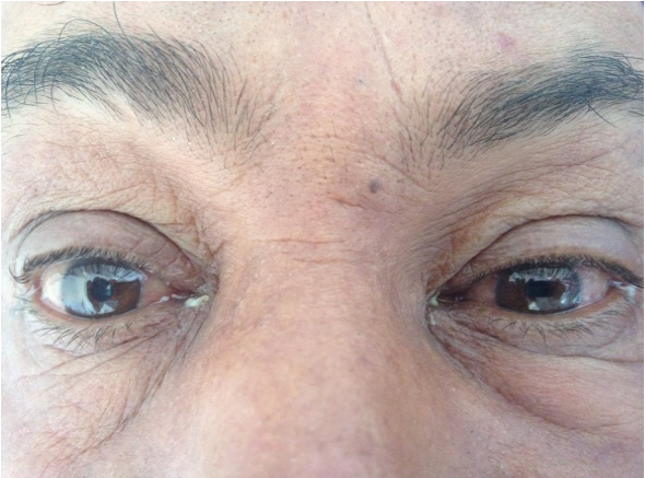
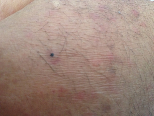
The somatic examination in our department noted a fever at 40°C, bilateral conjunctival hyperemia with signs of local irritation and tearing (Figure 1), maculopapular rash at both thighs and lower abdomen, and a small black necrotic lesion in the anterolateral aspect of the left thigh (typical "black spot" or “tache noire’’ of the MSF) (Figure 2).
The biological assessment revealed a marked inflammation with an erythrocyte sedimentation rate at 120mmH1, a C-reactive protein at 86mg/l and a polyclonal hypergammaglobulinemia at 19g/l. The blood count showed leukocytosis at 13 200/mm3 with 80% of neutrophils. The other basic biological tests were within normal limits: platelet, hemoglobin, creatinine, ionogram, glycaemia, liver enzymes, muscle enzymes, and urine analysis. Similarly, the chest x-ray, the electrocardiogram, and the abdominal ultrasound were without abnormalities.
The serodiagnosis of rickettsiosis was positive for Rickettsia conorii, confirming the diagnosis of MSF. The other specific investigations for infections (particularly tuberculosis), autoimmune diseases, neoplasms, and systemic affections were negative.
The patient was treated with oral doxycycline at a dose of 200mg/day. The evolution was rapidly favorable with apyrexia, total disappearance of the skin rash, of ocular complains, and progressive recuperation of vision. Ophthalmological exam with eye fundus was totally normal at two months, and without sequel.
Discussion
Ocular involvement during rickettsioses is often asymptomatic and spontaneously resolving, explaining its apparent rarity in clinical series [5].
These complications typically concern the retina and the optic disc, but all the tissues and parts of the eye can be affected [5,6]; it can be retinitis, retinal vascular involvement (occlusion, subocclusion, vascularitis, hemorrhage), endogenous endophthalmitis, optic disc edema, optic neuritis, neuroretinis, ischemic optic neuropathy, conjunctivitis, subconjunctival hemorrhage, keratitis, iris nodule, and oculomotor nerve palsies [5-8]. Ocular complications remain rare during MSF [7,9].
Uveitis is an exceptional and unusual ocular manifestation during rickettsial diseases, and rickettsioses are not cited among the classical infectious causes of uveitis [10,11]. Indeed, no case of rickettsiosis was found in the series of 2619 cases of uveitis of which 495 (19%) were of infectious causes [1].
Even in countries endemic for rickettsioses, uveitis is not usual. Indeed, only two cases of intermediate uveitis were related to rickettsiosis in the French series of 927 patients with uveitis of Bodaghi B., et al. (1.4%) [12], whereas no case was reported in the Turkish series of 761 cases of uveitis of Kazokoglu H., et al. [13].
Similarly in the Tunisian series of Khairallah M., et al. of 472 patients with uveitis, rickettsioses were involved in only five cases (1.1%) [14].
Uveitis associated with rickettsioses are classically posterior and bilateral, the anterior, intermediate, and unilateral forms are much rarer [7,15,16]. Total uveitis is exceptional and only a few sporadic cases are reported [7]. In fact, no case of rickettsial panuveitis was found in the series of 472 uveitis with 100 panuveitis of Khairallah M., et al. [14] nor in the series of 927 cases of uveitis with 324 total uveitis of Bodaghi B., et al. [12].
Thus rickettsioses are currently considered among the new etiologies of uveitis [17]; These also called "emergent" or "resurgent" diseases [1,3,17] are responsible of the new subgroup of unveite so-called "emergent infectious uveitis" [6]. The lack of knowledge and familiarity of most clinicians with these emergent etiologies of uveitis explains the diagnostic difficulties, and justifies the qualification of "inagmatic» given by some authors to ocular involvement in tick-borne infections [18].
This uveitis associated with rickettsioses can result from two pathophysiological mechanisms: direct inversion and bacterial proliferation in the cells of the ocular tissues (direct mechanism) or the immeasurable inflammatory and immunological reaction to the bacterial agent (indirect mechanism) [8].
The treatment consists of an appropriate oral antibiotic; doxycycline is the molecule of choice [7,8]. Glucosteroids are only indicated in severe forms and if immunological mechanism; indeed they can aggravate ocular involvement (especially if direct mechanism) [8]. Therefore an appropriate antibiotherapy is often indicated with the initiation of corticosteroid therapy in severe uveitis associated with rickettsial diseases.
Conclusion
A better knowledge of the rare causes of uveitis, especially the so-called emerging and reemerging infectious diseases, is necessary to take care of these affections adequately and in time. Only an early diagnosis of certainty and timely and appropriate management are the guarantors of a better functional ocular prognosis, since uveitis is the fourth most common cause of blindness in adults in developed countries.
The diagnosis of MSF should be evoked in front of any uveitis that does not prove or improve on corticosteroids, especially in endemic countries for rickettsioses.
Ethical considerations: Oral and written consent was given by patient for publication of his photos and data.
References
- Barisani-Asenbauer T., et al. “Uveitis- a rare disease often associated with systemic diseases and infections- a systematic review of 2619 patients”. Orphanet Journal of Rare Diseases 7 (2012): 57.
- Harman LE., et al. “Uveitis: the collaborative diagnostic evaluation”. American Family Physician 90.10 (2014): 711-716.
- Parola P., et al. “Update on tick-borne rickettsioses around the world: a geographic approach”. Clinical Microbiology Reviews 26.4 (2013): 657-702.
- Bouomrani S., et al. “Bilateral pleurisy revealing mediterranean spotted fever”. MOJ Clinical & Medical Case Reports 8.5 (2018): 189-191.
- Khairallah M., et al. “Novel infectious agents causing uveitis”. International Ophthalmology 30.5 (2010): 465-483.
- Khairallah M., et al. “Emergent infectious uveitis”. Middle East African Journal of Ophthalmology16.4 (2009): 225-238.
- Agahan AL., et al. “Intraocular inflammation as the main manifestation of Rickettsia conorii infection”. Clinical Ophthalmology 5 (2011): 1401-1407.
- Chawla R., et al. “Rickettsial retinitis: Direct bacterial infection or an immune-mediated response?” Indian Journal of Ophthalmology 65.10 (2017): 1038-1041.
- Verdot S., et al. “Ocular involvement in acute phase of Mediterranean boutonneuse fever”. Bull Soc Ophtalmol Fr 86.3 (1986): 429-431.
- Lin P. “Infectious Uveitis”. Current Ophthalmology Reports 3.3 (2015): 170-183.
- Tsirouki T., et al. “A Focus on the Epidemiology of Uveitis”. Ocular Immunology and Inflammation 26.1 (2018): 2-16.
- Bodaghi B., et al. “Chronic severe uveitis: etiology and visual outcome in 927 patients from a single center”. Medicine (Baltimore) 80.4 (2001): 263-270.
- Kazokoglu H., et al. “Demographic and clinical features of uveitis in tertiary centers in Turkey”. Ophthalmic Epidemiology 15.5 (2008): 285-293.
- Khairallah M., et al. “Pattern of uveitis in a referral centre in Tunisia, North Africa”. Eye (Lond) 21.1 (2007): 33-39.
- Belz., et al. “Fièvre boutonneuse et uvéite bilatérale”. Bull Soc Ophtalmol Fr 5 (1954): 429-431.
- Rizo AE., et al. “A case of bilateral uveitis secondary to Mediterranean spotted fever”. Clinical Infectious Diseases 14.2 (1992): 624-625.
- Khairallah M., et al. “New infectious etiologies for posterior uveitis”. Ophthalmic Research 49.2 (2013): 66-72.
- Argelich Ibáñez R., et al. “Enigmatic retinochoroiditis”. Med Clin (Barc) 140.9 (2013): e17.
Citation: Salem Bouomrani., et al. “Bilateral Panuveitis Revealing Mediterranean Spotted Fever”. Ophthalmology and Vision Science 3.1 (2019): 1-5.
Copyright: © 2019 Salem Bouomrani., et al. This is an open-access article distributed under the terms of the Creative Commons Attribution License, which permits unrestricted use, distribution, and reproduction in any medium, provided the original author and source are credited.


 Scientia Ricerca is licensed and content of this site is available under a Creative Commons Attribution 4.0 International License.