Editorial
Volume 2 Issue 2 - 2018
Intraosseous Rib Haemangioma
1Department of Orthopaedics, Hinduja Healthcare Surgicals, 11th Road, Khar (West), Mumbai
2Department of Orthopaedics, Bharati Hospital, Pune
3Department of Orthopaedics, Osmania General Hospital, Hyderabad
2Department of Orthopaedics, Bharati Hospital, Pune
3Department of Orthopaedics, Osmania General Hospital, Hyderabad
*Corresponding Author: Rohan Bhimani, Department of Orthopaedics, Hinduja Healthcare Surgicals, 11th Road, Khar (West), Mumbai.
Received: May 14, 2018; Published: May 23, 2018
Abstract
Introduction: The affliction of bone by Haemangioma is very uncommon and the involvement of ribs is extremely rare. Very few cases in literature have been reported on rib haemangiomas. This case report aims to highlight the presenting features of rib haemangioma and its management.
Case Report: A 23-year-old male presented with a chest x-ray demonstrating an incidental asymptomatic large expansile lesion on posterior aspect of the right 8th rib. On further evaluation by Computed tomography and biopsy the diagnosis of haemangioma was established. It was then managed by en-bloc surgical excision.
Conclusion: The diagnosis of haemangioma must be kept in mind when presented with cases of expansile lesion of rib. Rib haemangioma may show an aggressive nature on radiological investigations and they need to be confirmed and differentiated from primary bone malignant lesions of the rib by means of biopsy or excision.
Keywords: Haemangioma; Rib Tumour; Lytic lesions
Introduction
Haemangiomas account to about 1% of all bone neoplasms and frequently affects the skull and the vertebrae [1]. Rib involvement by haemangiomas are difficult to diagnose as they are usually seen at the bottom of differentials of rib lesions with handful of cases reported in literature [1-7]. We report a case of incidentally detected rib haemangioma in a 23-year-old male patient.
Case Report
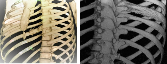
A 23-year-old male presented with a chest x-ray demonstrating an incidental asymptomatic large expansile lesion on posterior aspect of the right 8th rib. Computed Tomography (CT) on 500 slice dynamic 4D CT scanner was done. It showed posteriorly placed lesion of the right 8th rib involving its head and neck (Figure 1). It demonstrated an expansile, multiple, small irregular lucencies within and areas of cortical breaches. There was mild to moderate soft tissue involvement which was extrapleural in location with sun ray type spiculated periosteal reaction. No active pleuro-pulmonary pathology was seen in the scan (Figure 2).

Figure 2: Expansile, multiple, small irregular lucencies within and areas
of cortical breaches and spiculated sun ray type of periosteal reaction.
Further, CT guided biopsy was carried out. The biopsy report showed fibrinious material admixed with red blood cells and compactly arranged thin-walled blood capillaries lined by flattened endothelial cells. There was no evidence of malignancy and thus, the diagnosis of rib haemangioma was confirmed. As surgical resection of the affected rib was the treatment of choice, the excision through postero-lateral thoracotomy approach was carried out. The patient had an uneventful recovery and remained well at subsequent follow-ups. Microscopically, the specimen showed an ill-defined expansile lesion of the rib. The medullary component was replaced by soft, compactly packed, thin walled blood vessels with dilated channels. There were no cytological atypia. The diagnosis of benign haemangioma was established.
Discussion
Haemangioma is defined as a neoplastic entity which arises from blood vessels [3]. Even though skin is a common site of involvement, haemangioma of the bone comprises of about 1% of all bone tumours [1-7]. Pre-operative diagnosis of rib haemangioma is tenuous as most patients are asymptomatic and the discovery of lesion is incidental, as in our case. Plain roentogram is commonly inconclusive and various appearance like honeycomb, sun burst and soap bubble have been associated with rib haemangiomas in radiological diagnosis [4]. It is observed that asymptomatic bone lesions depicting aggressive nature, haemangioma should be considered. CT or MRI can delineate and identify the extent of involvement by the lesion. Haemangiomas do show a characteristic sun burst appearance with coarsed trabecular pattern on CT scan.
The histological diagnosis on an en-bloc specimen for haemangioma is not difficult and it gives definitive diagnosis. But, it may become catastrophic when biopsy is taken for confirmation as the tumour is very vascular and contains multiple arterial vessels. Histologically, haemangiomas are classified as cavernous, capillary, and venous or mixed depending on the type of vascular involvement [1]. The most common variety is cavernous haemangioma, constituting up to 50% of all cases [1]. This variety consists of large dilated vessels lined by a single layer of endothelial cells surrounded by fibrous stroma [2].
Since rib lesion includes multiple entities, the differential diagnosis plays a pivotal role. Fibrous dysplasia is the most common non-neoplastic tumour of the rib, accounts to 30% of benign bone tumours of the bony thorax. It classically appears as an expansile lytic area with ground glass appearance [6]. The chief treatment of haemangioma is surgical resection but large lesions usually carries the risk of bleeding profusely during surgery.
Thus, the diagnosis of haemangioma should be taken into consideration when dealing with cases of expansile rib lesion. In addition, a rib haemangioma may also demonstrate an aggressive nature on CT scan in form of cortical breach and periosteal reaction like sun burst appearance. CT guided biopsy or excision aids in diagnosis because a large portion of primary bone tumours are malignant. But, biopsy carries significant risk of bleeding. Thus, one needs to be prepared to manage when challenged by such complication.
Conflict of Interest
The authors declare that there is no conflict of interest regarding the publication of this paper.
The authors declare that there is no conflict of interest regarding the publication of this paper.
Data Availability
All data generated or analyzed during this study are included in this article.
All data generated or analyzed during this study are included in this article.
References
- Jain SK., et al. “Rib Haemangioma: A Rare Differential for Rib Tumours”. Indian Journal of Surgery 73.6 (2011): 447-49.
- Deshmukh H., et al. “Hemangioma of Rib: A Different Perspective”. Polish Journal of Radiology 80 (2015): 172-175.
- Okumura T., et al. “Hemangioma of the rib: a case report”. Japanese Journal of Clinical Oncology 30 (2000): 354-357.
- Narayan P., et al. “Rib haemangioma: A rarity and diagnostic dilemma”. Indian Journal of Thoracic and Cardiovascular Surgery 24.3 (2008): 212-214.
- Tew K., et al. “Intraosseous hemangioma of the rib mimicking an aggressive chest wall tumor”. Diagnostic and Interventional Radiology 17.12 (2011): 118-121.
- Ovali GY., et al. “Hemangioma of the Rib”. Turkish Journal of Medical Sciences 36.8 (2006): 65-70.
- Park JY., et al. “Cavernous Hemangioma of the Rib: A Case Report”. Iranian Journal of Radiology 13.3 (2016): e31677.
Citation:
Rohan Bhimani., et al. “Intraosseous Rib Haemangioma”. Chronicle of Medicine and Surgery 2.2 (2018): 129-131.
Copyright: © 2018 Rohan Bhimani., et al. This is an open-access article distributed under the terms of the Creative Commons Attribution License, which permits unrestricted use, distribution, and reproduction in any medium, provided the original author and source are credited.


 Scientia Ricerca is licensed and content of this site is available under a Creative Commons Attribution 4.0 International License.